
Descriptions
A miniaturized and low-power radiation camera equipped with particle tracking and imaging Timepix3 sensor (256 x 256 square pixels with a pitch of 55 μm). It offers versatility and portability for particle tracking and research.
Where can you use the MiniPIX MAGIC photon-counting camera? For material analysis or radiation monitoring.
- Spectral X-ray imaging: X-ray fluorescence imaging, X-ray radiography (low flux)
- Crystallography: Energy dispersive XRD, SAXS, or WAXS. We can analyse even thick samples or internal structures.
- Radiation monitoring: Particle type sorting, spectroscopy, directional sensitivity
- Particle physics research.
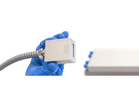
The MiniPIX MAGIC detector is connected via USB cable. The device has a microUSB port. The PIXet Pro software is provided with the device – it is used to operate the detector and collect and analyse data. Plugins for advanced functions are available.